
Skin Cancer Reconstruction Before & After Photos
Skin Cancer Reconstruction Before & After Case 1

Skin Cancer Reconstruction Before and After Photo. Surgery performed in Dallas, TX at Law Plastic Surgery.
Skin Cancer Reconstruction Before & After Case 2
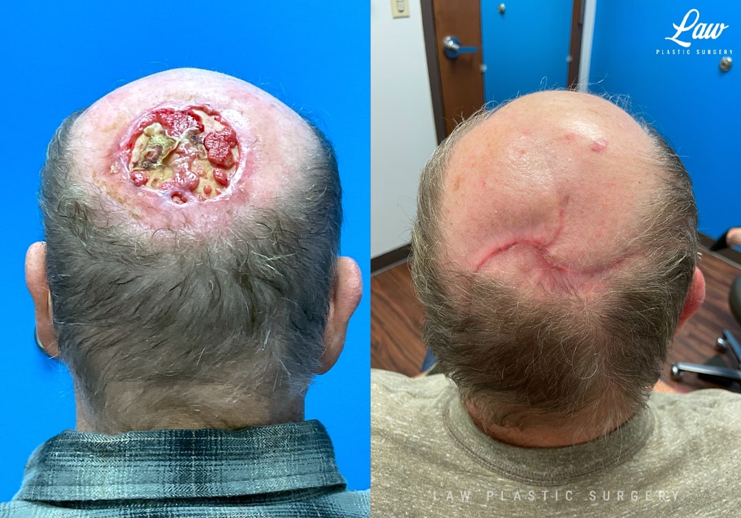
Reconstruction was performed with opposing rotation flaps, respecting the hair line and the blood flow to the scalp.
A few things I keep in mind for the reconstruction:
– I try to capture at least two primary blood vessels in each flap (from the supraorbital/supratrochlear, superficial temporal, postauricular, and occipital arteries),
– I plan to release widely but incrementally as needed,
– I will use subgaleal scoring as needed to help relieve tension,
– and I pay attention during the initial resection and the dissection to look for sources of blood flow to the margin of the wound.
Repair is performed with PDS suture in the galea for longer term support, and staples in the scalp maximize blood flow are well-tolerated.
He is seen at 4 months, with some redness from further topical treatments as directed by his dermatologist.
Skin Cancer Reconstruction Before and After Photo. Surgery performed in Dallas, TX at Law Plastic Surgery.
Skin Cancer Reconstruction Before & After Case 3
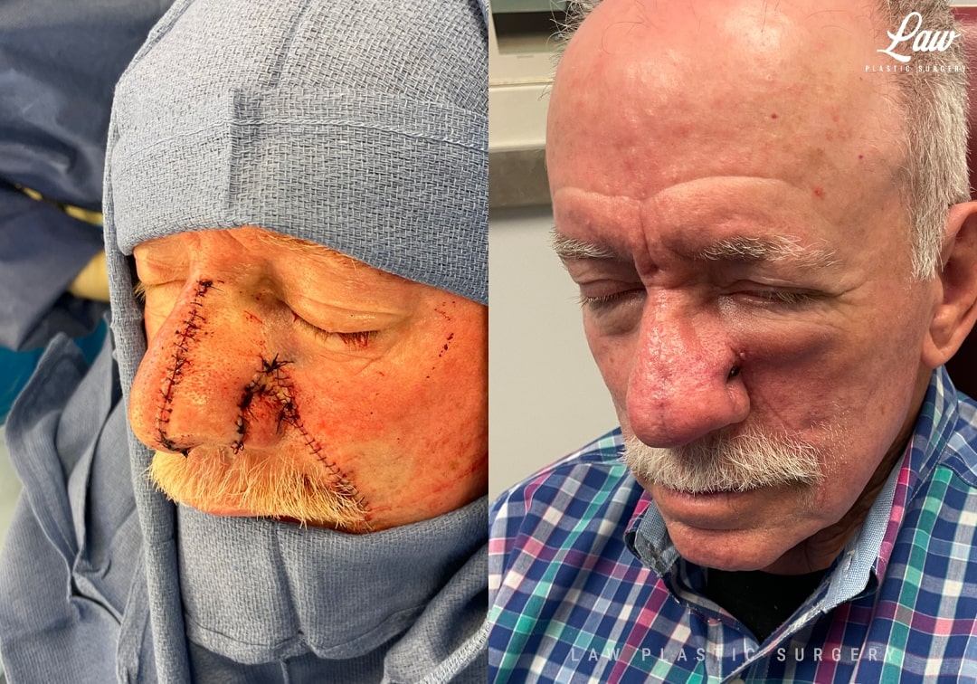
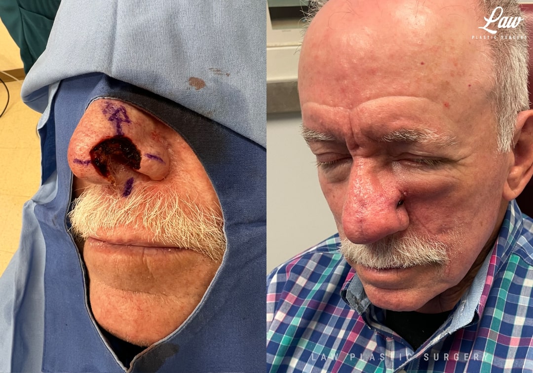
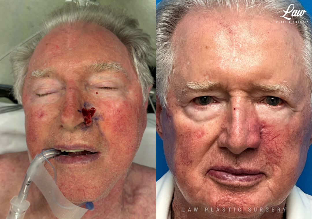
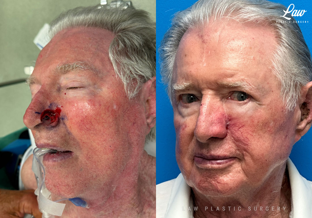
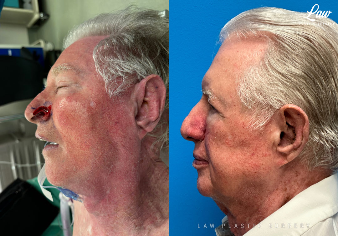
Here, a skin cancer involving the ala of the nose required full thickness excision, including the skin, cartilage, and nasal lining on the inside of the nostril.
A single-stage nasolabial flap provided the skin and nasal lining, and ear cartilage was used to provide structure and support.
Another option would be a two-stage forehead flap, where skin and muscle is taken from the forehead and rotated down. However, that requires 3-4 weeks with a bridge of skin from the forehead to the nose, and second surgery to divide and inset the flap. Sometimes, a third stage is even performed to improve the shaping and symmetry.
Results are seen here on the table results and at just 1 month, with the scars maturing nicely.
Skin Cancer Reconstruction Before and After Photo. Surgery performed in Dallas, TX at Law Plastic Surgery.
Skin Cancer Reconstruction Before and After Photo. Surgery performed in Dallas, TX at Law Plastic Surgery.
Skin Cancer Reconstruction Before and After Photo. Surgery performed in Dallas, TX at Law Plastic Surgery.
Skin Cancer Reconstruction Before & After Case 4
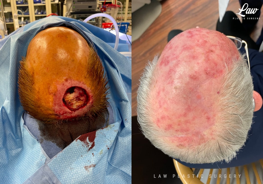
Here, a pinwheel design and release of areas of adhesion allowed the scalp to rotate and advance in to cover the exposed bone.
This was done as an outpatient going home the same day, without needing to take muscle or skin from another area of his body.
It also gave robust coverage that’s more durable than a skin substitute graft, and could be done in one stage instead of two or more.
He is seen here at 2 months healing up wonderfully.
Skin Cancer Reconstruction Before and After Photo. Surgery performed in Dallas, TX at Law Plastic Surgery.
Skin Cancer Reconstruction Before & After Case 5
Skin Cancer Reconstruction Before and After Photo. Surgery performed in Dallas, TX at Law Plastic Surgery.
Skin Cancer Reconstruction Before & After Case 6
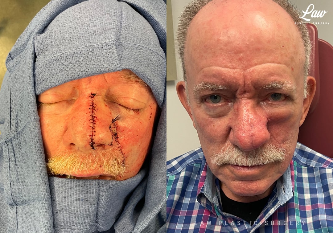
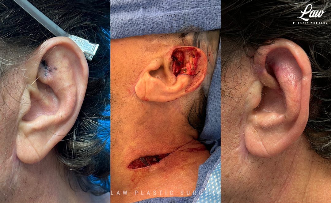
Traditionally, most surgeons would opt for a forehead flap. This involves building a bridge of skin from the forehead and eyebrow, to keep the skin on the nose alive while it starts healing in, and then dividing it a month later. For that month, the bridge can be unsightly, and can cause difficulty with visual obstruction, wearing glasses, and with wound care needs for the raw undersurface.
Here, I opted for a single-stage nasolabial flap from the left cheek crease. This type of flap can also be wrapped around the rim to resurface the lining on the inside of the nose.
A cartilage graft from the ear was used to provide support for the rim of the nose.
This is 3.5 weeks after surgery. Swelling continues going down over the next 6-18 months. If there is excess bulk (because the skin and fat from the cheek is thicker than the skin on the other side of the nose), we can do some contouring and thinning as a same-day surgery on his timeline between travel and activities.
Skin Cancer Reconstruction Before and After Photo. Surgery performed in Dallas, TX at Law Plastic Surgery.
Skin Cancer Reconstruction Before and After Photo. Surgery performed in Dallas, TX at Law Plastic Surgery.
Skin Cancer Reconstruction Before and After Photo. Surgery performed in Dallas, TX at Law Plastic Surgery.