
Ear Reconstruction Before & After Photos
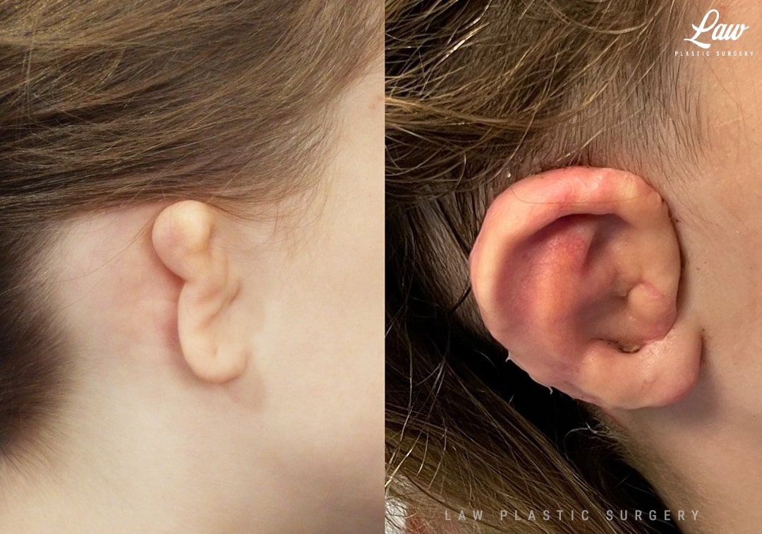
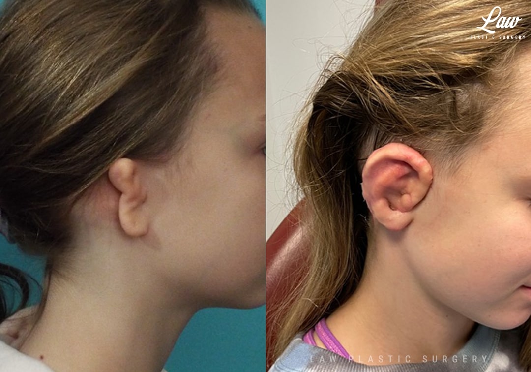
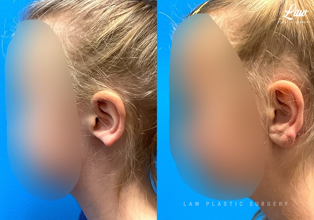
Ear Reconstruction Before & After 1
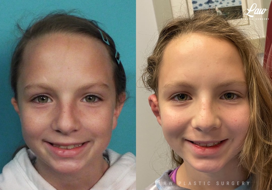
Ear Reconstruction Before and After Photo. Surgery performed in Dallas, TX at Law Plastic Surgery.
Ear Reconstruction Before and After Photo. Surgery performed in Dallas, TX at Law Plastic Surgery.
Ear Reconstruction Before and After Photo. Surgery performed in Dallas, TX at Law Plastic Surgery.
Microtia reconstruction is one of the most intricate and rewarding surgeries we perform as craniofacial surgeons.
Here, a modified Nagata technique was used to create a cartilage framework from the patient’s own ribs.
Guides and models based on the unaffected ear helped our design and placement of the new ear.
The first surgery involves the framework construction and placement under the skin (which is tailored and redraped).
At the second stage, support is placed behind the ear with cartilage saved from the first surgery. This provides projection away from the head, and local tissue and a skin graft are used to cover this area on the back of the ear.
There are pros and cons to this technique versus use of #medporearreconstruction.
A couple pros of this technique include more durability against infection, and no need for a TPF flap (and its scar, numbness, and possible hair loss).
The downsides include the rib donor site with a small risk of an air leak to the lungs, and need for two stages to achieve additional projection.
We are happily accepting new patients for new reconstructions, or revisions of previous reconstructions.
As always, photos are shared with specific written and verbal permission.
Ear Reconstruction Before & After 2
Ear Reconstruction Before and After Photo. Surgery performed in Dallas, TX at Law Plastic Surgery.
Ear Reconstruction Before and After Photo. Surgery performed in Dallas, TX at Law Plastic Surgery.
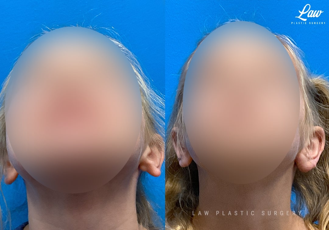
Ear Reconstruction Before and After Photo. Surgery performed in Dallas, TX at Law Plastic Surgery.
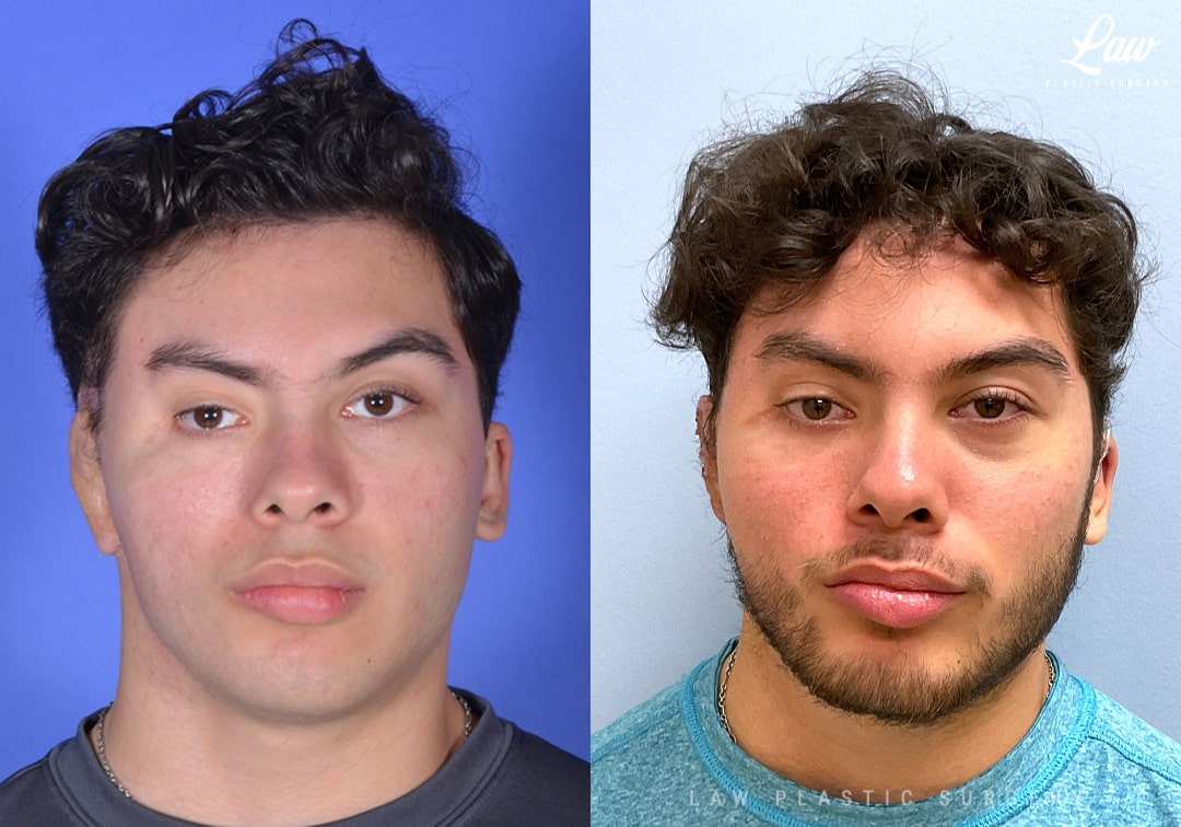
Differences in ear formation can come in all sorts of ways.
Here, this patient had an absent earlobe, with the rest of the structures of the ear, canal, and hearing, all unaffected.
Surgery was done to create an earlobe using a combination of cartilage for support and shape, and a flap of skin from behind her ear. This hid the donor site scar well, and gave a good match in color and texture.
Additional cartilage was placed on the inner side of the new lobe, buttressing the grafted cartilage to keep it from swinging forwards or backwards as easily as she heals and continues growing.
She is confident and happy, as we hope all of our patients will be as we get to see them mature and grow!
Ear Reconstruction Before & After 3
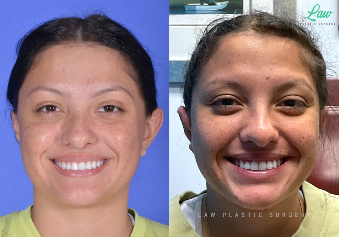
Ear Reconstruction Before and After Photo. Surgery performed in Dallas, TX at Law Plastic Surgery.
Ear Reconstruction Before and After Photo. Surgery performed in Dallas, TX at Law Plastic Surgery.
Ear Reconstruction Before and After Photo. Surgery performed in Dallas, TX at Law Plastic Surgery.
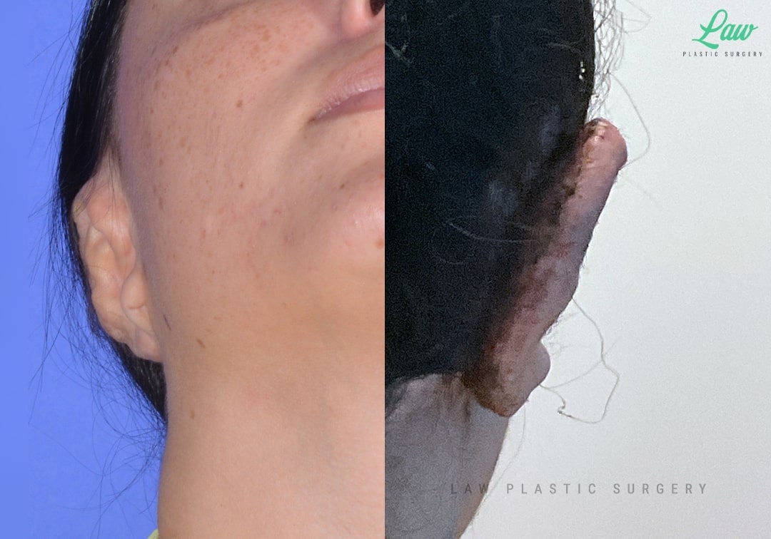
This lovely patient has a history of microtia and cartilage ear reconstruction done over 10 years ago.
She was happy with the result, but had difficulty wearing glasses because of the blunt angle at the top of the ear. She also had a hard time wearing masks or anything else that needs to loop around the ear because the lobe and crease behind the ear were also blunted.
We excised some of the old scar tissue and deepened the fold.
A full thickness skin graft from her old chest scar gave additional surface area to help change that flat slope into a deeper groove.
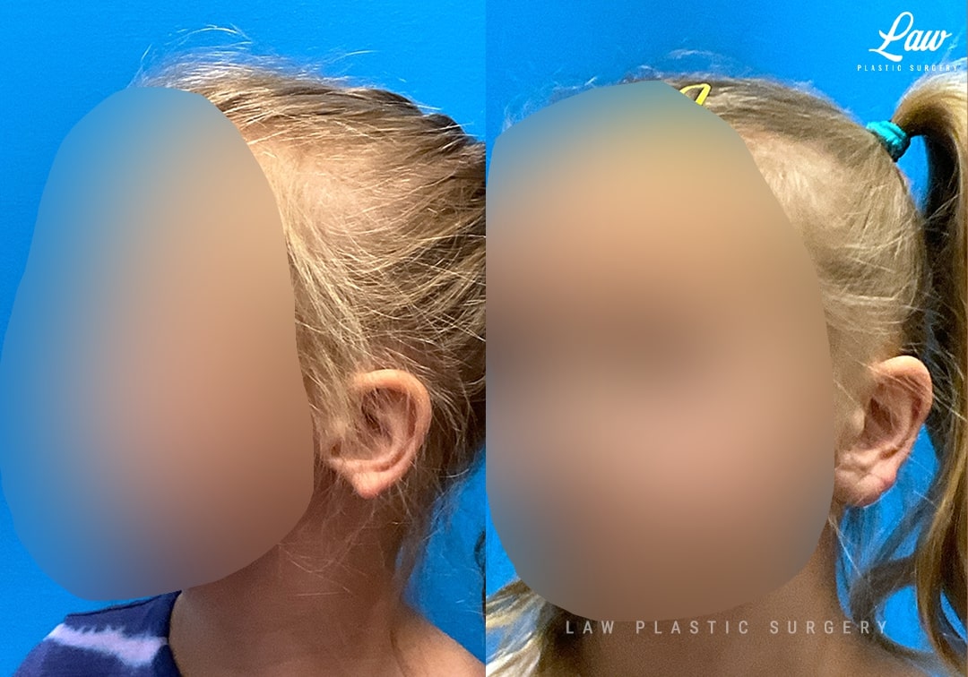
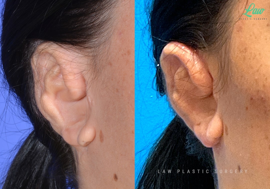
Ear Reconstruction Before & After 4
Ear Reconstruction Before and After Photo. Surgery performed in Dallas, TX at Law Plastic Surgery.
Ear Reconstruction Before and After Photo. Surgery performed in Dallas, TX at Law Plastic Surgery.

Ear Reconstruction Before and After Photo. Surgery performed in Dallas, TX at Law Plastic Surgery.
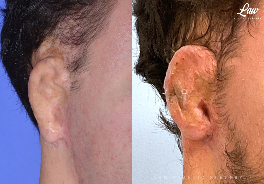
We have had several patients with previous ear reconstructions who have a hard time wearing a facemask or glasses.
The top of the ear can be blunted with a slope the doesn’t let anything stay on the ear.
Here, the ear was elevated and that depression deepened. A skin graft and advancement of the surrounding scalp was performed to add that depth.
The crease around the earlobe was also deepened to help with mask wearing. Finally, some rearrangement of the cartilage helped to round out the ear and add some depth to the contour.
This wonderful and always-optimistic patient is seen here at 3 weeks after surgery.